Home » Without Label » Anatomy Of Chest / Anatomy of thorax (2) : A detailed knowledge of chest wall anatomy is crucial for reconstructions in this difficult patient population.
Anatomy Of Chest / Anatomy of thorax (2) : A detailed knowledge of chest wall anatomy is crucial for reconstructions in this difficult patient population.
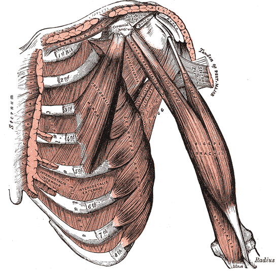
Anatomy Of Chest / Anatomy of thorax (2) : A detailed knowledge of chest wall anatomy is crucial for reconstructions in this difficult patient population.. Principal functions are the protection of internal viscera and an expandable cylinder facilitating variable gas flow into the lungs. A woman's chest — like the rest of her body — is covered with skin that has two layers. The dominant muscle in the upper chest is the pectoralis major. The right side of the heart is deflected anteriorly, and the left side is deflected posteriorly. You will also find the xiphoid process, 10th rib, the apex of the heart, the coronary vein of the heart.
Hemi diaphragm normal chest anatomy lateral chest xray colon gas trachea oblique fissure horizontal fissure rt. Anatomy of the thorax, heart, abdomen and pelvis recommended text gray's anatomy for students. The chest wall is comprised of skin, fat, muscles, and the thoracic skeleton. Plus, how to target each to make them bigger and stronger. The anatomic illustrations are presented as…
Trunk Muscles | Boundless Anatomy and Physiology from s3-us-west-2.amazonaws.com Chest a man's chest — like the rest of his body — is covered with skin that has two layers. The chest anatomy includes the pectoralis major, pectoralis minor and the serratus anterior. Plus, how to target each to make them bigger and stronger. This page provides an overview of the chest muscle group. A good radiologist knows the anatomy because knowing where structures normally live and recognizing the location of an abnormality helps to make or narrow the differential diagnosis. Anatomy of the thorax, heart, abdomen and pelvis recommended text gray's anatomy for students. It is important to remember the position and orientation of the heart when placing a stethoscope on the chest of a patient and listening for heart sounds, and also when looking at images taken from a midsagittal perspective. The pec major) is the one that commands the most real estate.
Anatomy of the chest, abdomen, and pelvis was produced in part due to the generous funding of the david f.
The chest anatomy includes the pectoralis major, pectoralis minor and the serratus anterior. This page provides an overview of the chest muscle group. The dominant muscle in the upper chest is the pectoralis major. 31 anatomy of the female breast syllabus p. The chest or thorax is the region between the neck and diaphragm that encloses organs, such as the heart, lungs, esophagus, trachea, and thoracic diaphragm. The epidermis is the outermost layer that provides a protective, waterproof seal over the body. Related posts of anatomy of the chest and stomach anatomy of pancreas. The right side of the heart is deflected anteriorly, and the left side is deflected posteriorly. The circulatory system does most of its work. Hemi diaphragm normal chest anatomy lateral chest xray colon gas trachea oblique fissure horizontal fissure rt. (1) the pectoralis major, and (2) the pectoralis minor. Principal functions are the protection of internal viscera and an expandable cylinder facilitating variable gas flow into the lungs. In insects, crustaceans, and the extinct trilobites, the thorax is one of the three main divisions of the creature's body, each of which is in turn composed of multiple segments.
A detailed knowledge of chest wall anatomy is crucial for reconstructions in this difficult patient population. The chest wall is comprised of skin, fat, muscles, and the thoracic skeleton. The first step in understanding thorax anatomy is to find out its boundaries. Large, complex chest wall defects can be some of the most challenging problems a reconstructive surgeon must face, but successful outcomes may be reliably achieved by adhering to basic principles of adequate debridement followed by. Principal functions are the protection of internal viscera and an expandable cylinder facilitating variable gas flow into the lungs.
Anatomy of the chest cavity — Medical Art Works from cdn.shopify.com 31 anatomy of the female breast syllabus p. Basic thoracic anatomy and physiology an understanding of thoracic imaging requires knowledge of the anatomy being imaged, as described in this chapter, as well as the imaging techniques applied to the thorax, covered in chapter 2. Thoracic cavity, also called chest cavity, the second largest hollow space of the body. A good radiologist knows the anatomy because knowing where structures normally live and recognizing the location of an abnormality helps to make or narrow the differential diagnosis. A line is drawn from anterior surface of the body of 6th thoracic vertebrae passing through the apex of the heart up to anterior lower most part of diaphragm. Related posts of anatomy of the chest area anatomy of male reproductive system. It is important to remember the position and orientation of the heart when placing a stethoscope on the chest of a patient and listening for heart sounds, and also when looking at images taken from a midsagittal perspective. Anatomy of the thorax, heart, abdomen and pelvis recommended text gray's anatomy for students.
Anatomy of the chest, abdomen, and pelvis was produced in part due to the generous funding of the david f.
Related posts of anatomy of the chest area anatomy of male reproductive system. 31 anatomy of the female breast syllabus p. Three dimensional view of the female reproductive system, full frontal view. Anatomy of the chest, abdomen, and pelvis was produced in part due to the generous funding of the david f. Anatomy of the thorax, heart, abdomen and pelvis recommended text gray's anatomy for students. 2 skin of the anterior chest wall syllabus p. Sternocleidomastoid muscle clavicle and ribs anatomy muscle anatomy chest sternocleidomastoid ribs anatomy chest muscles anatomy thorax rib muscles chest muscles chest anatomy illustration. Related posts of anatomy of the chest and stomach anatomy of pancreas. The anatomic illustrations are presented as… This page provides an overview of the chest muscle group. In insects, crustaceans, and the extinct trilobites, the thorax is one of the three main divisions of the creature's body, each of which is in turn composed of multiple segments. A good radiologist knows the anatomy because knowing where structures normally live and recognizing the location of an abnormality helps to make or narrow the differential diagnosis. Browse 6,407 chest anatomy stock photos and images available, or search for human anatomy to find more great stock photos and pictures.
Anatomy of the thorax, heart, abdomen and pelvis recommended text gray's anatomy for students. Basic thoracic anatomy and physiology an understanding of thoracic imaging requires knowledge of the anatomy being imaged, as described in this chapter, as well as the imaging techniques applied to the thorax, covered in chapter 2. The epidermis is the outermost layer that provides a protective, waterproof seal over the body. Computed tomography (ct) of the chest can detect pathology that may not show up on a conventional chest radiograph(1). The right side of the heart is deflected anteriorly, and the left side is deflected posteriorly.
Chest anatomy, artwork - Stock Image - F005/9996 - Science ... from media.sciencephoto.com (1) the pectoralis major, and (2) the pectoralis minor. About the 6th week, the somites differentiate into the sclerotomes and the dermatomyotomes. Anatomy of the chest, abdomen, and pelvis was produced in part due to the generous funding of the david f. Sternocleidomastoid muscle clavicle and ribs anatomy muscle anatomy chest sternocleidomastoid ribs anatomy chest muscles anatomy thorax rib muscles chest muscles chest anatomy illustration. The chest wall, like other regional anatomy, is a remarkable fusion of form and function. Knowledge of the anatomy of the whole cylinder (ribs, sternum, vertebra, diap … 2 skin of the anterior chest wall syllabus p. 4 innervation of the breast blood supply of the breast syllabus p.
The chest wall, like other regional anatomy, is a remarkable fusion of form and function.
2 skin of the anterior chest wall syllabus p. The human thorax includes the thoracic cavity and the thoracic wall. Chest a man's chest — like the rest of his body — is covered with skin that has two layers. 31 anatomy of the female breast syllabus p. The anatomic illustrations are presented as… Large, complex chest wall defects can be some of the most challenging problems a reconstructive surgeon must face, but successful outcomes may be reliably achieved by adhering to basic principles of adequate debridement followed by. The chest is made up primarily of two muscles: A good radiologist knows the anatomy because knowing where structures normally live and recognizing the location of an abnormality helps to make or narrow the differential diagnosis. Here, we break down the anatomy of your chest muscles. Anatomy of pancreas 12 photos of the anatomy of pancreas anatomy and histology of pancreas ppt, anatomy and physiology of human pancreas, anatomy of pancreas divisum, describe the microscopic anatomy of pancreas, gross anatomy of pancreas, human anatomy, anatomy and histology of pancreas ppt, anatomy and physiology of. The chest anatomy includes the pectoralis major, pectoralis minor and the serratus anterior. About the 6th week, the somites differentiate into the sclerotomes and the dermatomyotomes. See human chest anatomy stock video clips.